

Application Notes
Imaging Technology Applied Products: Prokaryote or eukaryote? A unique microorganism from the deep sea
Imaging Technology Applied Products : Prokaryote or eukaryote? A unique microorganism from the deep sea
Prokaryote or eukaryote? A unique microorganism from the deep sea
- Authors (Members in bold)
Masashi Yamaguchi1*, Yuko Mori2, Yoshimichi Kozuka3, Hitoshi Okada1,4, Katsuyuki Uematsu5, Akihiro Tame5, Hiromitsu Furukawa2, Tadashi Maruyama6, Cedric O’Driscoll Worman7 and Koji Yokoyama1
1Medical Mycology Research Center, Chiba University, 2System in Frontier Inc., 3Vacuum Device Co. Ltd., 4Integrated Imaging Research, 5Marine Works, 6Marine Biodiversity Research Program, Japan Agency for Marine-Earth Science and Technology, 7Department of Biology, Francis Marion University
- Published
- Journal of Electron Microscopy, Volume 61, Issue 6, December 2012, Pages 423‒431,
https://doi.org/10.1093/jmicro/dfs062
- Abstract
- There are only two kinds of organisms on the Earth: prokaryotes and eukaryotes. Although eukaryotes are considered to have evolved from prokaryotes, there were no previously known intermediate forms
between them. The differences in their cellular structures are so vast that the problem of how eukaryotes could have evolved from prokaryotes is one of the greatest enigmas in biology. Here, we report a unique organism with cellular structures appearing to have intermediate features between prokaryotes and eukaryotes, which was discovered in the deep sea
off the coast of Japan using electron microscopy and structome analysis. The organism was 10 μm long and 3 μm in diameter, having >100 times the volume of Escherichia coli. It had a large ‘nucleoid’, consisting of naked DNA fibers, with a single nucleoid membrane and endosymbionts that resemble bacteria, but no mitochondria. Because this organism appears to be a life form distinct from both prokaryotes and eukaryotes but similar to eukaryotes, we named this unique microorganism the ‘Myojin parakaryote’ with the scientific name of Parakaryon myojinensis (‘next to (eu)karyote from Myojin’) after the discovery location and its intermediate morphology. The existence of this organism is an indication of a potential evolutionary path between prokaryotes and eukaryotes.
- Abstract
- There are only two kinds of organisms on the Earth: prokaryotes and eukaryotes. Although eukaryotes are considered to have evolved from prokaryotes, there were no previously known intermediate forms between them. The differences in their cellular structures are so vast that the problem of how eukaryotes could have evolved from prokaryotes is one of the greatest enigmas in biology. Here, we report a unique organism with cellular structures appearing to have intermediate features between prokaryotes and eukaryotes, which was discovered in the deep sea off the coast of Japan using electron microscopy and structome analysis. The organism was 10 μm long and 3 μm in diameter, having >100 times the volume of Escherichia coli. It had a large ‘nucleoid’, consisting of naked DNA fibers, with a single nucleoid membrane and endosymbionts that resemble bacteria, but no mitochondria. Because this organism appears to be a life form distinct from both prokaryotes and eukaryotes but similar to eukaryotes, we named this unique microorganism the ‘Myojin parakaryote’ with the scientific name of Parakaryon myojinensis (‘next to (eu)karyote from Myojin’) after the discovery location and its intermediate morphology. The existence of this organism is an indication of a potential evolutionary path between prokaryotes and eukaryotes.
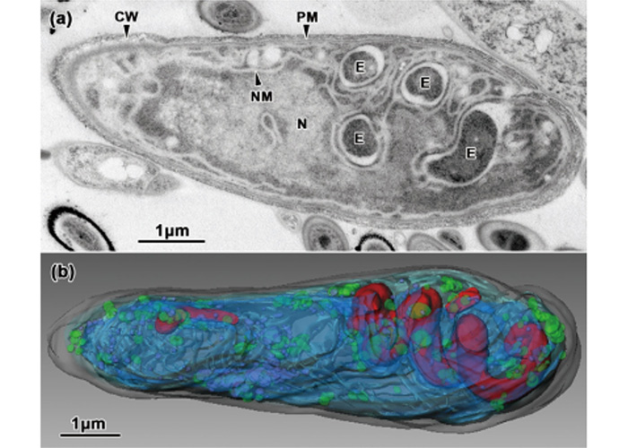
(b)3D reconstruction results from our application.
In addition, in a joint research with Dr. Yamaguchi of Chiba University, the following papers have been published.
- Title
- High-voltage electron microscopy tomography and structome analysis of unique spiral bacteria from the deep sea
- Authors (Members in bold)
Masashi Yamaguchi, Hiroyuki Yamada, Kimitaka Higuchi, Yuta Yamamoto, Shigeo Arai, Kazuyoshi Murata, Yuko Mori, Hiromitsu Furukawa, Mohammad Shorif Uddin, and Hiroji Chibana
- Published
Microscopy, 2016, 1‒7
https://doi.org/10.1093/jmicro/dfw016
- Title
- Good Ultrastructural Preservation of Human Tissues and Cultured Cells by Glutaraldehyde Fixation, Sandwich Freezing, and Freeze-Substitution
- Authors (Members in bold)
Masashi Yamaguchi, Seiichiro Wakabayashi, Yuumi Nakamura, Hiroyuki Matsue, Takuya Hirao, Shigeki Aoki, Shohei Yamashina, Hiroyuki Yamad, Nobuya Mamizu, Hiromitsu Furukawa and Hiroji Chibana
- Published
Cytologia 2020 85(1): 15-26
https://doi.org/10.1508/cytologia.85.15